Skip to content
Monday, July 14, 2025
About
Download Report
CSR
Contact Us
North City Diagnostic Center
Think Health Think NorthCity
Advanced Technologies
Departments
Health Packages
In House Doctor
Health Info
Systemic Diseases
Infective Diseases
Organ Specific Diseases
Health Tips
Bookings
Vacancies
site mode button
Search for:
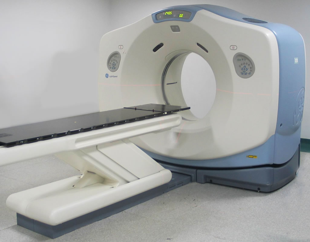
CT SCAN
Equipments
August 17, 2024
August 17, 2024
admin
Post navigation
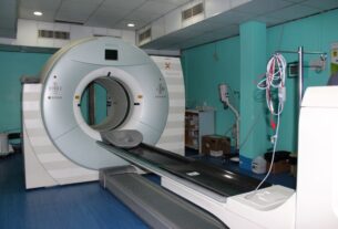
PET CT
Access 2
Related Posts
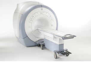
MRI
August 18, 2024
August 18, 2024
admin
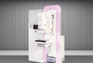
Mam Venus DRV
August 17, 2024
admin
PET CT
August 17, 2024
admin