

Our Diagnostic Services
Topmost Quality. Reliable Results. 27 yrs
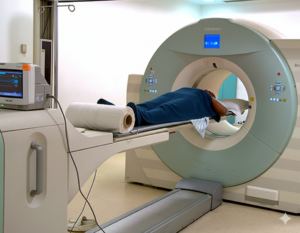
PET CT(128 Slice)
FDG/PSMA/DOTANOC PET
A positron emission tomography (PET) scan is an imaging test that creates images of your organs and tissues at work. The test uses a safe, injectable radioactive chemical called a radiotracer and a device called a PET scanner. The scanner detects cells that absorb large amounts of the radiotracer, which indicates a potential health problem. A PET scan can measure vital functions, such as blood flow, oxygen use and metabolism.
A healthcare provider may order a PET scan to check for signs of:
- Cancer, including breast cancer, lung cancer and thyroid cancer
- Coronary artery disease, heart attack or other heart problems
- Brain disorders, such as brain tumors, epilepsy, dementia and Alzheimer’s disease
- Determine if cancer has spread, evaluate if cancer treatment is working or see if cancer has returned
- Evaluate damage from a heart attack or stroke and identify areas of your heart that need treatment
They can also use it to:
- Phone:9230993800
- Email:web.booking@northcitydiagnostic.com
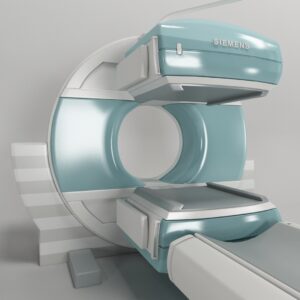
Gamma Camera
BONE SCAN/DTPA/DMSA/THYROID SCAN
A whole-body bone scan is a test to check the health of your bones. Healthcare providers typically order this scan to check whether cancer has spread (metastasized) to your bones. You may also have a whole-body bone scan to help determine cancer stage (how advanced it is).
A whole-body bone scan is a type of nuclear medicine imaging scan. Providers use advanced technology that may sound complicated. But the tests are painless, effective and safe for most people.
Before the test, you receive an injection of a radiotracer (a substance that contains a small, safe amount of radioactive material). The radiotracer collects in areas of your bones where there are changes or irregularities. These areas appear brightly on an imaging scan, helping your provider pinpoint disease.
What does a whole-body bone scan show?
A whole-body bone scan can show damage or changes in your bones. The test doesn’t show your provider the cause of the damage or changes.
Your provider may use a whole-body nuclear bone scan as one of several tools when diagnosing conditions like:
- Avascular necrosis.
- Bone cancer.
- Bone fractures.
- Bone infections (osteomyelitis).
- Fibrous dysplasia.
- Osteoarthritis.
- Paget’s disease of the bone (osteitis deformans).
- Unexplained bone pain.
- Phone:9230993800
- Email:web.booking@northcitydiagnostic.com
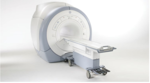
MRI (1.5T)
MRI Brain/Spine/ Angio/Spectro/MRCP
An MRI (magnetic resonance imaging) scan is a painless test that produces very clear images of the organs and structures inside your body. MRI uses a large magnet, radio waves and a computer to produce these detailed images. It doesn’t use X-rays (radiation).
Because MRI doesn’t use X-rays or other radiation, it’s the imaging test of choice when people will need frequent imaging for diagnosis or treatment monitoring, especially of their brain.
What does an MRI show?
Magnetic resonance imaging (MRI) produces detailed images of the inside of your body. Healthcare providers can “look at” and evaluate several different structures inside your body using MRI, including:
- Your brain and surrounding nerve tissue.
- Organs in your chest and abdomen, including your heart, liver, biliary tract, kidneys, spleen, bowel, pancreas and adrenal glands.
- Breast tissue.
- Your spine and spinal cord.
- Pelvic organs, including your bladder and reproductive organs (uterus and ovaries in females and the prostate gland in males).
- Blood vessels.
- Lymph nodes.
- Phone:9230993800
- Email:web.booking@northcitydiagnostic.com
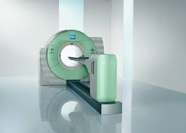
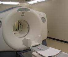
CT SCAN
CT Brain/Chest/ Abdomen/CT Angio
A CT (computed tomography) or CAT (computed axial tomography) scan is a type of imaging test that helps detect diseases and injuries. It uses X-rays and a special computer to create detailed pictures of your bones, organs and soft tissues.
It creates images of the inside of your body by taking multiple X-ray pictures at different angles. When the images are combined, it creates a 3D image. This lets your provider see your body very clearly and in much more detail than a standard X-ray.
CT scans can show if a health condition is getting better or worse with treatment. It can detect things such as cancer and certain infections. What does a CT scan show?
CT scans can show abnormalities like tumors, injuries and diseases in more detail than an X-ray. Examples include:
- Many types of cancer
- Pneumonia and emphysema
- Broken bones
- Heart disease
- Blood clots
- Bowel disorders (appendicitis, diverticulitis, blockages, Crohn’s disease)
- Kidney stones
- Brain injuries
- Spinal cord injuries
- Internal bleeding
- Phone:9230993800
- Email:web.booking@northcitydiagnostic.com

Cardiac CT (128 Slice)
CT Coronary Angio/ CT Calcium Scoring
A cardiac computed tomography (CT) scan is a procedure that utilizes multiple X-ray beams from different angles to acquire high-quality, three-dimensional (3D) images of your heart, along with your great vessels and surrounding structures.
Cardiac CT uses advanced CT technology, with or without intravenous (IV) contrast (dye) to better visualize your heart structure and associated blood vessels. With multi-slice scanning, your healthcare provider can get high-resolution, 3D images of your moving heart and great vessels.
What does a CT scan of the heart show?
Your healthcare provider will be able to see your:
- Coronary arteries that supply your heart.
- Heart chambers, muscle and valves.
- Pulmonary veins.
- Thoracic aorta, and sometimes abdominal aorta.
- Sac around your heart (pericardium).
- Phone:9230993800
- Email:web.booking@northcitydiagnostic.com

USG
Abdomen/Pregnancy/Anomaly/Elastography
Ultrasound (also called sonography or ultrasonography) is a noninvasive imaging test that lets healthcare providers “see” inside your body. Ultrasound uses high-frequency sound waves to create real-time pictures or video of internal organs or other soft tissues. An ultrasound picture is called a sonogram. A doctor or a healthcare provider called an ultrasound technologist or sonographer performs ultrasounds.
Healthcare providers use ultrasound to look at different parts of your body for many reasons.
The three main categories of ultrasound are:
- Pregnancy ultrasound: Providers use prenatal ultrasound to confirm you’re pregnant, estimate your due date and check fetal growth.
- Diagnostic ultrasound: Providers use diagnostic ultrasounds to view internal organs and tissues to see if something is wrong or not working properly. Examples include an abdominal ultrasound, breast ultrasound and transrectal ultrasound.
- Ultrasound guidance for procedures: Providers sometimes use ultrasound to help guide needles, catheters or other instruments during a surgery or procedure. For example, a biopsy or nerve block can require ultrasound guidance.
3D and 4D ultrasounds
- Most ultrasound images are 2D (two-dimensional). This is the most common type of ultrasound imaging healthcare providers use. It produces a flat image of your organs and internal structures.
- Three-dimensional (3D) ultrasound is another type of ultrasound. This type produces more detailed images because it shows the depth and angles that a 2D ultrasound can’t produce.
- Four-dimensional (4D) ultrasound is 3D ultrasound in motion. You can think of it as a “live” 3D image.
- Phone:9230993800
- Email:web.booking@northcitydiagnostic.com
Gastroenterology
Endoscopy & Colonoscopy
Endoscopic ultrasound (EUS) is a procedure to examine the inside of your digestive tract. A gastroenterologist (a doctor who specializes in the digestive system) performs an EUS.
An EUS test combines:
- Endoscope, a long, thin, flexible tube with a camera and light on the end.
- Ultrasound, a probe on the end of the endoscope that sends out sound waves to produce images of your organs, tissues and blood vessels.
- Abdominal (belly) pain or chest pain.
- Cancer spread (metastatic cancer).
- Lumps, lesions (abnormal tissue) or growths seen on previous imaging scans like CT scans or MRIs.
- Digestive tract problems, such as inflammatory bowel disease (IBD), colon cancer and anal fissures.
- Gallbladder diseases, including gallstones and cholecystitis (gallbladder inflammation).
- Lung diseases, such as lung cancer.
- Pancreatic diseases, such as pancreatic cancer, pancreatic cysts and pancreatitis.
- Swallowing disorders like Barrett’s esophagus.
Who needs an endoscopic ultrasound (EUS) procedure?
Healthcare providers use EUS to evaluate or diagnose:
- Lining of the esophagus, stomach and small intestine.
- Lymph nodes, blood vessels, cysts and tumors.
- Nearby organs, such as the pancreas, gallbladder and bile ducts.
What are the types of EUS tests?
An upper endoscopy procedure is an EUS procedure that examines the upper part of the digestive tract, including:
- Anal sphincter (muscles surrounding the anus).
- Blood vessels, lymph nodes, cysts and tumors.
- Lining of the large intestine (rectum and colon).
A lower EUS procedure examines the lower part of your digestive tract. This includes:
- Phone:92309933800
- Email:web.booking@northcitydiagnostic.com
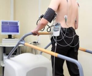
Cardiology
ECG/ECHO/TMT/PFT
An electrophysiology study (EP study) is a detailed analysis of the electrical activity in your heart. Electrophysiology (EP) is a subject within cardiology that analyzes your heart’s electrical signals to diagnose and treat abnormal heart rhythms (arrhythmias).
Your healthcare provider uses cardiac catheters (small tubes) and computers to make electrocardiogram (EKG) tracings and electrical measurements from inside your heart. Sensors a provider places inside your heart provide more information than you can get from electrodes outside your chest.
Studying your heart’s electrical activity can tell your provider if and where something is going wrong with your heartbeat signals. Normally, signals that tell your heart to beat travel the same route in an organized way every time.
When you have an abnormal heart rhythm, it’s like a bus that isn’t following its normal route. The bus may start the route at the wrong bus stop, skip a stop or not travel the full route. Or it could be going too fast or too slow. Irregular signals can be like this. Your provider wants to find out why to determine whether the rhythm is dangerous and how to treat it.
What is electrophysiology used for?
Your EP study can give your healthcare provider information about your abnormal heart rhythm, such as:
- What kind of arrhythmia you have.
- What’s causing your abnormal heart rhythm.
- What part of your heart is causing the issue.
- Why you’ve been feeling dizzy or fainting.
- Where in your heart an abnormal rhythm starts.
- Which treatment is best for you.
- How well your medicine is working.
- Whether you may be at risk for sudden cardiac death.
- A standard EKG.
- Holter monitor.
- Event recorder.
- Stress test.
- Echocardiogram.
- Angiogram.
Your provider may want to do an EP study when other tests can’t provide enough information about your abnormal heart rhythm.
You may have already tried these tests:
Depending on your condition, an electrophysiologist can use the information from an EP study to treat you with catheter ablation right away. They can do this through the catheters already in place in your heart for the study.
- Phone:92309933800
- Email:web.booking@northcitydiagnostic.com
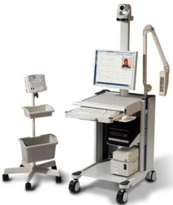
Neurology
EEG/EMG/NCV
A neurological (Neuro) exam consists of a physical examination to identify signs of disorders affecting your brain, spinal cord and nerves (nervous system). Neurological examination is the best way for healthcare providers to check the function of your brain and nervous system. It helps them determine which tests to run. Every neurological exam is different depending on your symptoms. Sometimes, it can take time to complete the physical examination and the interview, as your nervous system is very complex.
If you already live with a nervous system disorder, a neurological exam helps healthcare providers track treatment response.
What does a neuro exam entail?
It includes a series of physical examination steps that assess nervous system functioning. These may include evaluation of your:
- Mental status (cognition) and speech.
- Cranial nerve (nerves of your head and face) function.
- Strength, coordination and muscle tone.
- Reflexes (such as “knee jerk”) and others.
- Perception of feeling in different parts of your body to different stimulation, like touch and vibration.
- Gait and mobility.
- Spine.
- Level of consciousness, after a traumatic brain injury, for example.
- Altered state of consciousness, including coma.
- Cognitive decline, including confusion, memory loss or changes in behavior.
- Blurred or double vision, impaired hearing or loss of sense of smell.
- Difficulty with speech (dysarthria or aphasia).
- Difficulty with balance or coordination.
- Dizziness.
- Headaches.
- Muscle weakness.
- Numbness or tingling in your arms and legs.
- Seizures.
Who may need a neurological exam?
You may benefit from a neurological exam if you have symptoms of neurological dysfunction such as:
- Phone:92309933800
- Email:web.booking@northcitydiagnostic.com
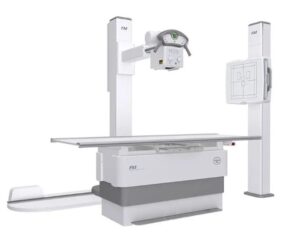
Digital X Ray
X Ray CHEST/JOINT/BONES
An X-ray is a type of medical imaging that uses radiation to take pictures of the inside of your body. We often think of an X-ray as something that checks for broken bones. But X-ray images can help providers diagnose other injuries and diseases, too.
Many people think of X-rays as black-and-white, two-dimensional images. But modern X-ray technology is often combined with other technologies to make more advanced types of images.
X-rays can help healthcare providers diagnose various conditions in your body. Some of the most common areas on your body to get an X-ray are:
Abdominal X-ray: This X-ray helps providers evaluate parts of your digestive system and diagnose conditions like kidney stones and bladder stones.
Bone X-ray: You might get a bone X-ray if your provider suspects you have a broken bone, dislocated joint or arthritis. Images from bone X-rays can also show signs of bone cancer or infection.
Chest X-ray: Your provider might order a chest X-ray if you have symptoms like chest pain, shortness of breath or a cough. It can look for signs of infection in your lungs or congestive heart failure.
Head X-ray: These can help providers see skull fractures from head injuries or conditions that affect how the bones in your skull form, like craniosynostosis.
Spine X-ray: A provider can use a spine X-ray to look for arthritis and scoliosis.
- Phone:92309933800
- Email:web.booking@northcitydiagnostic.com
Mammography
Mammo Single/ Both Breast
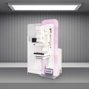
A mammogram is a low-dose X-ray of your breast tissue. Healthcare providers use mammograms, or mammography, to look for early signs of breast cancer before symptoms develop. This is called a screening mammogram. Providers also use mammography to look for any abnormalities if you develop a new symptom, such as a lump, pain, nipple discharge or breast skin changes. This is called a diagnostic mammogram.
Aside from skin cancer, breast cancer is the most common cancer that affects females and represents 15% of all new cancer diagnoses in the United States. An early diagnosis plays a big role in your breast cancer survival rate. Routine mammograms are key to catching breast cancer early. When breast cancer is detected early, the five-year survival rate is 99%. Mammograms are the gold standard in detecting breast tissue changes before they turn into cancer.
What are the different types of mammograms?
There are two main types of mammograms:
- Digital mammography in 2D.
- Digital mammography in 3D (digital breast tomosynthesis).
2D mammograms
- 2D mammography involves taking pictures of each breast at two different angles — typically from top to bottom and side to side.
3D mammograms
- 3D mammography is a newer type of mammogram in which each breast is compressed once and a machine takes several X-rays as it moves in an arc over your breast. A computer then puts the images together in “slices,” which allows healthcare providers to see your breast tissues in 3D.
- Phone:92309933800
- Email:web.booking@northcitydiagnostic.com

DENTAL
OPG/CBCT
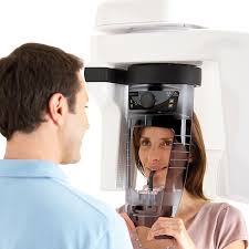
Dental X-rays (radiographs) are images of your teeth and jaws. Dentists use them to see parts inside that aren’t visible during a regular exam — like your jawbone, nerves, sinuses and tooth roots. A dental X-ray machine captures these images during your visit.
What can dental radiographs detect?
Dental X-rays help your dentist find a wide range of oral health issues, including:
- Cavities, especially between teeth
- Decay under existing fillings
- Bone loss in your jaw
- Unerupted or impacted teeth
- Abscesses (infections at the tooth root or between gums and teeth)
- Cysts and some tumors
Dentists also use X-rays to determine if you’re a candidate for treatments like implants, braces or dentures. X-rays help track healing after procedures like bone grafts or root canals.
- Phone:92309933800
- Email:web.booking@northcitydiagnostic.com
Get to the venue
Dedicated to providing affordable diagnostic services
REACH US
Get Direction to Diagnostic Centre
35A, Canal West Road, Near Gouribari , Kolkata 700004
North City Diagnostic Centre Pvt. Ltd.
35A, Canal W Rd, Gouri Bari, Shyam Bazar, Kolkata, West Bengal 700004

