Menu
Gallium PET (GA 68)
DOTATOC and PSMA Scan - Prominent images for finer diagnosis

In recent times, PET CT studies have gained much importance in detecting many diseases. In many diseases like neuroendocrine tumors, prostate malignancies etc. are often missed on a standard FDG PET study. Ga-68 labeled PET studies are asked for such clinical situations. Alike standard PET scans, the G8-Ga labeled PET studies help in diagnosing a tumor, help in characterizing the same, helps in staging-restaging of the disease etc.
Ga-68 is a radiotracer that emits positron and helps in doing PET scans. Ga-68 can be tagged with various components, namely DOTATOC and PSMA, depending upon the clinical need.
Ga-68 delivers prominent images for finer diagnosis
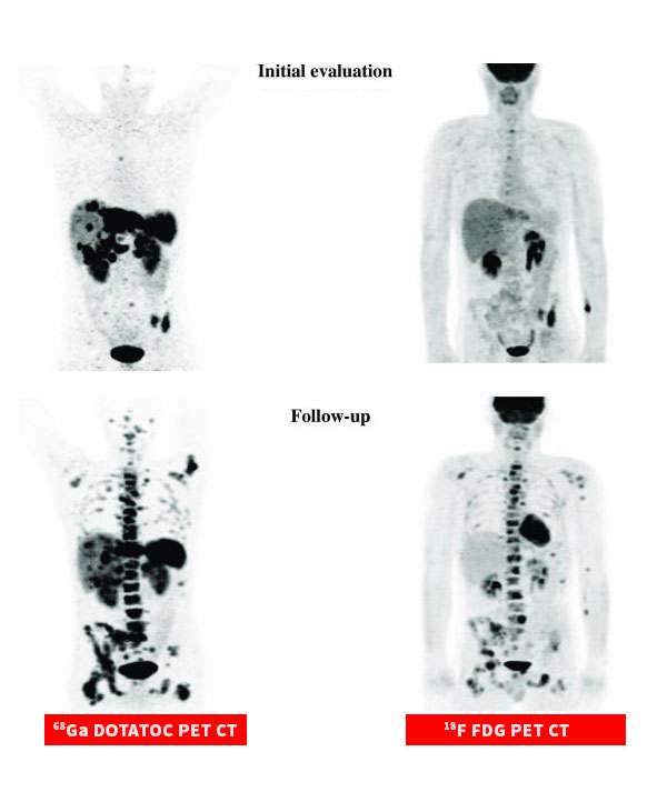
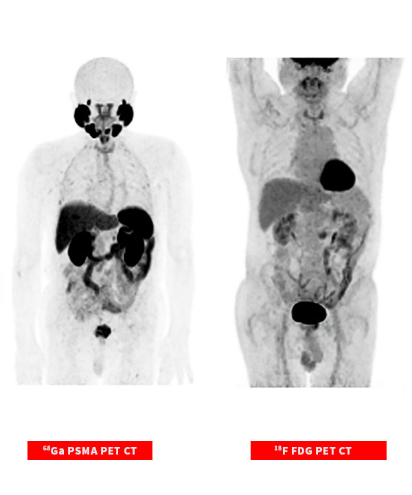
Capturing incidences that often goes undetected in a standard FDG PET CT Study
DOTATOC PET CT Scan
It is a scan to find and look at somatostatin receptor positive neuroendocrine tumors (NETs) in adults and kids. Ga-68 DOTANOC PET/CT scan is a whole-body examination which gives information regarding the site and extent of primary NET and distant metastasis, along with, somatostatin – receptor expression in individual lesions.
The Ga-68 labeled radiopharmacy is intravenously injected and a after waiting period (depending on the scan type) the PET CT scan is performed.
There are no major risk with radiopharmacy. A detailed consent would however be obtained before the procedure.
Although carcinoid tumors usually have good prognosis, early and specific diagnosis is important. Computed tomography and magnetic resonance imaging do not provide findings that are specific for carcinoids, and somatostatin receptor scintigraphy suffers from low spatial resolution. 18-Fluorodeoxyglucose positron emission tomography/computed tomography (18F-FDG PET/CT) has limited sensitivity for carcinoids due to low uptake of the marker.
Studies have shown °Ga-DOTATOC have much higher detection rate, highly accurate in the diagnosis of neuroendocrine tumors, meningiomas, etc. It has also been used successfully to evaluate primary lung cancers, particularly the small-cell form. The high resolution of PET enables high detection rates for small tumors. It is very is useful in the evaluation of primary pulmonary carcinoids and may be included in the diagnostic work-up of these patients.
PSMA PET CT Scan
PSMA PET imaging is a scan with more precise detection of prostate cancer for better treatment planning and targeted care. It is more effective in pinpointing and delineating tumors not only in the prostate but also throughout the body in cases where the tumors have migrated.
“Prostate Specific Membrane Antigen (PSMA) PET Imaging for Prostate Cancer Offering breakthrough diagnostics for prostate cancer treatment”
- PSMA PET imaging is a scan with more precise detection of prostate cancer for better treatment planning and targeted care.
- Imaging with PSMA PET was able to detect significantly more prostate lesions than conventional imaging who had undergone a radical prostatectomy but had experienced a recurrence of their cancer.
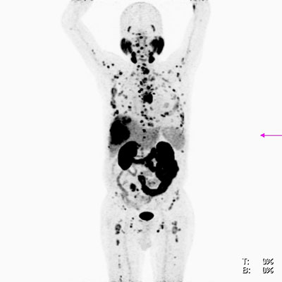
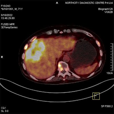
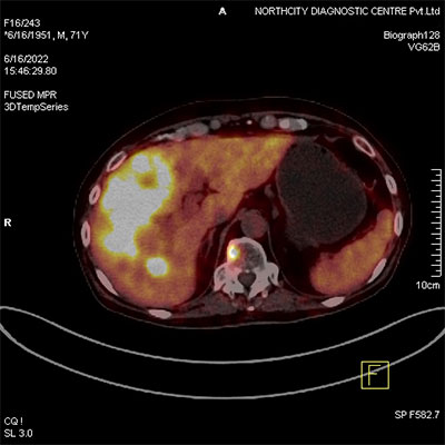
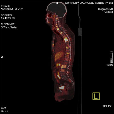
PSMA PET CT study at North City Diagnostic Centre
The PET CT study shows multiple PSMA expressing hypodense lesions in liver and multiple PSMA expressing patchy sclerotic lesions in bones – suggestive of metastatic disease.
Why North City Diagnostic Centre?
- 25 years old organization.
- NABL accredited lab for more than 12 years.
- State-of-the-Art technologies and diagnostic facilities all under one roof.
- Online Booking & Reporting facilities
- Reports prepared by highly experienced team of doctors, pathologists, radiologists and biochemists


