Magnetic Resonance Imaging (MRI)
Fast Facts on MRI Scanning
- MRI scanning is a non-invasive and painless procedure.
- Raymond Damadian created the first MRI full-body scanner, which he nicknamed the Indomitable.
An MRI scan uses a large magnet, radio waves, and a computer to create a detailed, cross-sectional image of internal organs and structures.
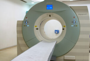
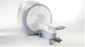
The scanner itself typically resembles a large tube with a table in the middle, allowing the patient to slide in.
An MRI scan differs from CT scans and X-rays, as it does not use potentially harmful ionizing radiation.
Uses
The development of the MRI scan represents a huge milestone for the medical world.
Doctors, scientists, and researchers are now able to examine the inside of the human body in high detail using a non-invasive tool.
The following are examples in which an MRI scanner would be used:
- anomalies of the brain and spinal cord
- tumors, cysts, and other anomalies in various parts of the body
- breast cancer screening for women who face a high risk of breast cancer
- injuries or abnormalities of the joints, such as the back and knee
- certain types of heart problems
- diseases of the liver and other abdominal organs
- the evaluation of pelvic pain in women, with causes including fibroids and endometriosis
- suspected uterine anomalies in women undergoing evaluation for infertility
- This list is by no means exhaustive. The use of MRI technology is always expanding in scope and use.