

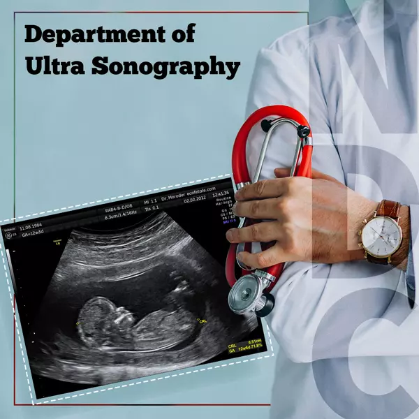
Ultra Sonography (USG) incorporates non-invasive procedures that use high-energy sound waves to look at tissues and organs inside the body. The sound waves make echoes that form pictures of the tissues and organs on a computer screen, called sonogram. It is used to investigate abdominal and pelvic organs, musculoskeletal and vascular systems. The development of fetus during pregnancy is monitored with USG.
An ultrasound scan or Sonogram, commonly called Ultrasonogram (USG)is used to examine internal body structures.
Ultrasound imaging sends out (emits) high-frequency sound waves, directed at the tissue being examined, and recording the reflected sound or echoes to create an image.
An ultrasound scan is generally non-invasive.
Common reasons for ultrasound scanning include investigations of a person’s abdominal and pelvic organs, musculoskeletal and vascular systems, and to check fetal development during pregnancy.
There are several types of ultrasound tests. Each uses a probe designed to image specific areas of the body. An abdominal ultrasound shows organs and other soft tissues (such as blood vessels) inside your abdomen (belly).
For an abdominal ultrasound test, a trained medical professional (sonographer) applies a special gel to your belly. The sonographer then moves the probe over the gel.
Sound waves from the probe go through your skin and bounce back from soft tissues (such as organs). Real-time (live) images show up on a computer screen nearby.
Your provider orders ultrasound evaluation of specific areas of your abdomen. A right upper quadrant ultrasound examines three organs of the digestive system:
A complete abdominal ultrasound examines those three organs and adds the:
Abdominal ultrasound is a common test to check on an unborn baby throughout pregnancy. Providers often call this test a prenatal ultrasound.
Abdominal ultrasound may also help pinpoint the cause of unexplained abdomen (stomach) pain. This test aids in the diagnosis of many routine problems (such as kidney stones) and more serious health concerns (such as blood clots).
Providers use abdominal ultrasound tests to detect:
A member of US will give you complete instructions before your exam. Follow their instructions to ensure the most accurate test results.
We may ask you to stop eating some hours before your test in cases of USG Upper of Abdomen. In cases of USG Lower Abdomen, you may need to drink a specific amount of water right before your test. In case of USG Whole abdomen you may need fasting and plenty of liquid intake. In some other cases you may not need to prepare at all.
Transvaginal Ultrasound, also called an endovaginal ultrasound, is a type of pelvic ultrasound used by doctors to examine female reproductive organs. This includes the uterus, fallopian tubes, ovaries, cervix, and vagina.
Unlike a regular abdominal or pelvic ultrasound, where the ultrasound wand (transducer) rests on the outside of the pelvis, this procedure involves your doctor or a technician inserting an ultrasound probe about 2 or 3 inches into your vaginal canal.
There are many reasons why a transvaginal ultrasound might be necessary, including:
Your doctor might also recommend a transvaginal ultrasound during pregnancy to:
A technique called ElastPQ uses ultrasound shear wave elastography (ARFI technology) to provide a noninvasive, reproducible, and easily performed method of assessing liver fibrosis. A special pulse sequence technique that uses existing transducers produces shear waves in tissue and then measures the propagation speed of the waves. Now liver stiffness samples can be acquired during a routine ultrasound examination of the liver. According to a recent study, using shear wave elastography may help reduce or avoid conventional liver biopsies.
Diffuse liver disease is one of the major health problems in the world. It can result from many causes, including viral hepatitis (hepatitis B or hepatitis C), non-alcoholic or alcoholic fatty liver disease, autoimmune hepatitis, drug-induced liver injury, primary biliary cirrhosis, and several other less frequent etiologies. It is estimated that 360 million and 180 million people worldwide are infected with viral hepatitis B and C respectively. Between 500,000 and 700,000 people die annually as a result of hepatitis B virus infection, and more than 350,000 people are estimated to die each year from hepatitis C-related liver disease.1-3 Chronic liver damage results in hepatic fibrosis, characterized by an increase in extracellular matrix material produced by fibroblast-like cells. This process results in liver fibrosis that can progress to cirrhosis with distortion of normal liver architecture and portal hypertension.
According to a recent study, using shear wave elastography may help reduce or avoid conventional liver biopsies.
With increasing fibrosis, the liver becomes stiffer, which can be monitored using shear wave elastography.13,14 With this technique, during an ultrasound exam after selecting the best acoustic window, a region of interest (ROI) is placed in an area of the liver perpendicular to the liver capsule, taking care not to include large vasculature or biliary structures. An intercostal imaging approach targeting segments 7 or 8 of the liver has been shown to provide more reliable measurements.
FibroScans do not require any special preparation, although you may be asked not to eat or drink for a few hours before the test. The procedure is completely painless, requires no sedation, and only takes a few minutes.
In Trans Rectal Sonography, the rectum is injected with a probe that emits high-energy sound waves. The sound waves are bounced off internal tissues or organs and make echoes. Echoes create a picture of body tissue called a sonogram. During the transrectal ultrasound, abnormalities in the prostate and nearby structures are screened for. ERUS and TRUS are other terms for the endorectal ultrasound.
Transrectal ultrasounds may cause a minimal amount of discomfort similar to what happens during a rectal exam. If our practitioner does a biopsy during the ultrasound, you may experience a more intense sensation in the rectum each time they retrieve a sample.
Prior to TRUS, the patient may be instructed to have an enema to remove feces and gas from the rectum, which might impede the progress of the rectal probe
The patient traditionally lies on his left side, which is considered a more relaxing position as well as allowing for easier insertion of the rectal probe. After the probe is inserted into the rectum, the tester adjusts the console on the ultrasound machine to a baseline for the echoes of normal prostate tissue, which will serve as the standard by which other tissue will be classified. Imaging usually starts at the base of the bladder, as the probe is rotated to provide a full picture of the prostate.
Most men in the age group for prostate cancer usually have some BPH as well, which can elevate PSA levels and make prostate cancer diagnosis more difficult. PSA density — the blood PSA level divided by the size of the prostate, as determined by TRUS — can help distinguish between BPH and prostate cancer.
With BPH, the PSA level should not be more than 15 percent of the size of the prostate. PSA levels exceeding 15 percent of the size of the prostate are more likely to indicate the presence of prostate cancer, and the need for a biopsy.
35-A ,Canal West Road
Near Gouri Bari Bus Stop
Kolkata – 700004.
Local: +91 33 6605 0888