


Imagine a time not too long ago when detecting the presence and progression of cancer or other critical illnesses was akin to solving a complex puzzle with many missing pieces. Patients had to go through multiple imaging tests, waiting anxiously for results, and the uncertainty of the diagnosis felt like an endless ordeal.
Today we have the technology for precision imaging. It is the PET CT Scans. The PET CT scan accuracy provides a clear picture of the patient’s condition, allowing doctors to devise a precise treatment plan. This technological breakthrough has transformed diagnostic imaging, bringing clarity and confidence to both patients and healthcare providers.
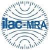
A PET CT scan is a hybrid imaging technique that combines Positron Emission Tomography (PET) with Computed Tomography (CT). The PET scan detects metabolic activity by tracing a radioactive substance injected into the body, while the CT scan provides detailed anatomical images. Together, these scans offer comprehensive insights into both the structure and function of tissues and organs.
PET CT scans play a crucial role in diagnosing various conditions, including cancer, heart disease, and neurological disorders. By highlighting areas of abnormal metabolic activity, they enable doctors to identify disease at an early stage, monitor the effectiveness of treatments, and detect recurrences.
The capabilities of PET CT scans are vast. They can:
The field of PET CT imaging has seen significant advancements, continually enhancing its accuracy, efficiency, and diagnostic capabilities. Here are some of the key advancements:
Modern PET CT scanners offer significantly improved image resolution and sensitivity. Advances in detector technology, such as digital PET detectors, have led to clearer images and better detection of small lesions. These improvements help in early diagnosis and more precise disease progression and treatment response monitoring.
Time-of-Flight (TOF) PET technology measures the exact time difference between the detection of the two gamma photons emitted during positron annihilation. This information is used to improve image quality by increasing the signal-to-noise ratio, making the images sharper and more accurate. TOF PET is particularly beneficial for imaging larger patients where image quality can be challenging.
Integrating PET with advanced CT technologies, such as dual-energy CT or spectral CT, provides additional diagnostic information. Dual-energy CT can differentiate materials based on their energy absorption characteristics, which can help in characterizing tissue types and identifying specific diseases.
Artificial intelligence (AI) and machine learning algorithms are being increasingly applied to PET CT imaging. These technologies assist in image reconstruction, noise reduction, and automated interpretation, enhancing diagnostic accuracy and reducing the time needed for analysis. AI can also help in predicting treatment outcomes and personalizing patient care.
New hybrid imaging modalities combining PET with MRI (PET/MRI) have emerged, offering the metabolic insights of PET with the superior soft tissue contrast of MRI. This combination is particularly valuable in neuroimaging, oncology, and cardiac imaging, where detailed anatomical and functional information is critical.
The development of novel radiotracers has expanded the scope of PET CT imaging. These tracers target specific biological pathways and receptors, providing more precise and targeted imaging. For instance, tracers targeting prostate-specific membrane antigen (PSMA) are used for detecting prostate cancer with high sensitivity.
Personalized dosimetry ensures that the amount of radioactive tracer used is tailored to the individual patient’s needs, optimizing image quality while minimizing radiation exposure. This approach enhances patient safety and improves diagnostic accuracy.
Integrating PET with advanced CT technologies, such as dual-energy CT or spectral CT, provides additional diagnostic information. Dual-energy CT can differentiate materials based on their energy absorption characteristics, which can help in characterizing tissue types and identifying specific diseases.
Gallium-68 (Ga-68) is a positron-emitting radionuclide widely used in PET CT imaging, particularly in oncology. Here’s what makes Ga-68 significant:
Gallium-68 is a radioactive isotope of gallium, commonly used in the form of Gallium-68-labeled radiopharmaceuticals. It is produced in a generator and has a short half-life of about 68 minutes, which makes it suitable for clinical PET imaging.
The accuracy of PET CT scans is one of their most significant advantages. They are highly sensitive and can detect abnormalities at the cellular level, often before structural changes are visible on other imaging modalities. This early detection capability is vital for effective treatment planning.
PET CT is considered part of nuclear medicine because it involves the use of radioactive tracers to visualize metabolic processes within the body. The radioactive substance emits positrons, which are detected by the PET scanner, creating detailed images of cellular activity. This nuclear medicine approach allows for precise diagnosis and treatment planning.
Dos
While PET CT scans involve radiation exposure, the benefits often outweigh the risks, especially for diagnosing and monitoring serious conditions like cancer. However, there is a general recommendation to limit unnecessary exposure. The frequency of scans is typically determined by medical necessity, and doctors ensure that the benefits justify the radiation exposure.
The side effects of PET CT scans are generally minimal. The radioactive tracer is usually well-tolerated, with rare instances of allergic reactions. Some patients may experience minor discomfort at the injection site or feel anxious about the procedure, but serious complications are uncommon.
When comparing 18F-FDG PET and PET Ga-68, it’s essential to understand their unique strengths to make an informed decision.
Your doctor’s recommendation will be based on the specific type of cancer and the diagnostic detail needed. Both methods are effective, but choosing the right one depends on your specific medical condition.
At North City Diagnostic Center we offer personalized testing schedule that aligns with your medical history and risk factors. Consistent monitoring can be invaluable for early detection and effective management of arthritis. It’s not just about how often you test but making sure that the tests are aligned with your overall health profile for maximum benefit.
#PETCTScanAccuracy #MRI #CT #Ultrasound #MedicalImaging #Diagnostics #Healthcare #NDC #HealthFirst #NorthCity #DiagnosticServices #Ga68 #CancerImaging #Oncology #NuclearMedicine #PSMA #DOTATOC
35-A ,Canal West Road
Near Gouri Bari Bus Stop
Kolkata – 700004.
Local: +91 33 6605 0888


